
About Common Lumps Bumps
Any localized swollen areas on or under the skin can be described as lumps and lumps. They can happen on the facial skin as well as other parts of the body. There are many causes, but people can be scared of the possibility of a cancer. Many of these can be diagnosed by simple inspection and the doctor can tell whether it is benign or potentially malignant. Some of these will resolve spontaneously, some are best left alone, while some can be simply removed or require the skills of a plastic surgeon.
Melanocytic Naevi (Moles)
Naevi are extremely common and all individuals have such blemishes on the skin. Some consult for cosmetic reasons or because they worry about their nature. Naevi are benign growth of any cells normally present in the skin. So there are a great varieties of them. An epidermal naevus is a benign growth of keratinocytes, and a melanocytic naevus is a benign growth of melanocytes, which is commonly referred to as a mole.
Melanocytic naevi can be present at birth or acquired in later life. There are different types of acquired melanocytic naevi:
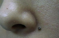
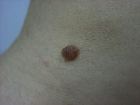
Junctional naevus: the naevus cells are located at the junction between the epidermis and dermis. It is flat, appears brownish black.
Intradermal naevus: the naevus cells are located in the dermis only. They are raised and mostly flesh-coloured, although some can be pigmented.
Compound naevus: a mixture of junctional and intradermal proliferation. They are slightly raised and brownish black.
Blue naevus: it is blue in colour as the melanocytes are very deep in the dermis.
 |
 |
Melanocytic naevi are related to sun exposure and they are therefore much more common in fair-skinned individuals, while individuals with darker skin types might have fewer naevi because of the protective properties of melanin from sunlight exposure. They may also darken following sun exposure or during pregnancy
Melanocytic naevi are benign but can be found in association with melanoma. The true frequency of transforming into melanoma is not known. If there is any doubt about the appearance, particularly when there is a change of size, thickness, darkness etc, always consult a dermatologist who will be able to tell if there is any need for further investigation.
Those clinically benign naevi can be removed by simple excision but this is going to need suture with the result of a scar, which is cosmetically unacceptable, particularly on the face. The raised intradermal types can be dealt with by shaving using an electric cautery. Now the CO2 laser is used to remove any of these easily, leaving only a mark, which will subside in a matter of months, and no trace afterwards.
Sebaceous Cysts
It is also called epidermoid cysts. Cysts are non-cancerous closed pockets of tissue that can be filled with fluid, pus, or other material. It is a benign skin cyst formed from blocked sebaceous glands in the skin. Apart from the face, it is most commonly found on the trunk, back, and scalp.
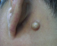
A little black opening of the cyst is visible on the skin. If it is infected, it will become red and tender. When it is squeezed, a cheesy white paste material is discharged, sometimes can be quite smelly.
To remove it effectively, the whole sac of the cyst must be completely removed. If the cyst is squeezed to force out the discharge, or if part of the sac wall is left behind, the cyst will reappear later.
Skin Tags
Skin tags are very common. The medical term is fibro-epithelial polyp. They typically occur around the neck, underarms, eyelids, groins and under the breasts, where the skin rubs against skin or clothing. They are benign skin growth. Each looks like a small, oval, hanging piece of skin with a narrow stalk, and can be flesh-coloured or pigmented.
They are more common in older people; after pregnancy; and in over-weight people who are more likely to have skin friction or rubbing.
Skin tags are harmless and will not turn into cancer. Occasionally they may get irritated and bleed; or snagged by clothing or jewelry and cause pain. They are just a piece of redundant skin and can be removed very easily.
Small ones can be cut off with scissors with no anesthesia. Freezing using liquid nitrogen, and burning using an electric cautery are both effective methods to remove larger ones, sometimes under a small amount of local anaesthesia.
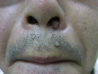
Plane Warts
All warts are infection of the skin surface by the human papilloma viruses. Plane warts appear as fleshed-coloured or pigmented, very slightly raised, well-defined and flat-topped lumps.
They are particularly common on the face and hands. When they are small they are not very noticeable, but with time they will spread and become larger, and numerous of these seem to appear suddenly and can be cosmetically alarming. They can be freezed off with liquid nitrogen, burn off with an electric cautery or CO2 laser.
Common Warts
Common warts are larger, round lumps with an uneven surface like that of a cauliflower. It is very common on the hands and fingers, and it frequently spreads to other parts like the face by touching and scratching. Cases have been seen with warts on any parts of the face, including the nostrils.
 |
 |
Treatment is similar to that of plane warts.
Seborrhoeic Keratosis
Seborrhoeic keratosis is caused by excessive growth of the top layer of epidermis. It is related to skin aging and sun exposure, is usually found in people over 30 years old, and tends to become more common and more numerous with age.
It is commonly seen on the face and scalp, but also often found on the chest and back. It can be solitary or in clusters. It appears as a lump of light tan to black colour, with sizes range from a few mm to a few cm, and has a look of a rough lump being stuck onto the surface.
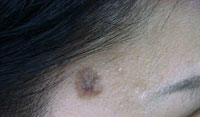
It is unsightly, especially when appearing on the face, and in a cluster of numerous lumps. It can get irritated by friction and scratching. It may grow much larger over the years and removal is sometimes recommended not just for cosmetic reason, especially if they get irritated and bleed easily. It is easily removed by scrapping off with the based cauterized afterwards, or the whole thing can be burn off by a CO2 laser, or freezed off using liquid nitrogen. Occasionally it may turn black and become difficult to distinguish from a skin cancer, in which case removing it intact for examination is mandatory.
Lipoma
Lipoma is a benign growth of the subcutaneous fat tissue. It tends to form on the trunk, shoulders, neck, but can appear elsewhere on the body and appears as solitary nodule or in groups. It has a firm, rubbery consistency. It is usually asymptomatic, but it can cause pain when it compresses nerves around it when it grow big enough.
Lipoma does not need to be removed unless there is a cosmetic concern, a compression of surrounding structures, or an uncertain diagnosis. If necessary, it can be removed easily by excision.
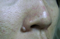
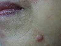
Sebaceous Gland Hyperplasia
Hyperplasia is an overgrowth of cells. Sebaceous gland hyperplasia is the overgrowth of the cells lining the sebaceous glands (oil-producing glands). It appears as a round, yellowish or fleshed-coloured lump, with a central depression, mostly on the face. It is associated with exposure to androgen hormones and sun, and is seen more commonly in men than in women.
It is a harmless condition and is only a cosmetic concern. It can be treated effectively by electric cautery or CO2 laser.