
Tinea
This is a superficial infection of the skin or nail with fungi.
What Are Fungi?
Fungi are microorganisms different from bacteria or viruses. They are present widely in nature. They can be separated into Dermatophytes and Yeasts. They are contracted from three major sources: soil, animals, and humans. Yeasts can exist naturally in our body, for example the Pityrosporum Ovale on the skin that causes seborrhoeic dermatitis, and Candida live inside the bowel and vagina.
Factors that may increase the likelihood of infection:
- Poor nutrition
- Poor personal hygiene
- Wet and humid weather
- Contact with infected animals
- Contact with infected persons
- Impaired personal immunity
Signs
Fungi normally invade just the superficial skin layer called stratum corneum. Different signs appear when different part of the body is infected.
Signs on the skin include:
- Itchy rash
- Dry and thickened skin
- Blisters
- Scaling and peeling
Clinical Patterns
Tinea PedisThis is often referred to as athlete’s foot, and is one of the commonest tinea infection of the skin. 30 - 70 % of people have tinea pedis at anytime of their life. This is normally a problem of the adults, only rarely seen in children. This is seen most frequently in tropical regions, particularly during the hot summer or wet and humid seasons.
The fungi causing the tinea pedis can be present inside shoes, socks, or on the floor, especially in public area such as bathrooms, or swimming pools. Too tight and occlusive shoes and socks, or excessive sweating of the feet, are also major contribution factors. Fungi can continue to live on clothing that has been washed. However, simple contact with this or infected skin scales is not enough to transmit the infection, there has to be a minor skin break that allows for the entry before infection will occur.
There are few different patterns in the infection.
Wet type:Mostly seen in the toe webs, and usually between the 3rd and 4th, or the 4th and 5th toes. The skin appears wet and soggy, sometimes macerated and has a bad smell. It is itchy and can be painful. If the skin is secondarily infected with bacteria, it will be red, swollen, and filled with pus.
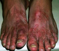
The skin on the sole becomes dry, hard, and thickened. It is red but there are some white flaky skin peeling off. The area can extend to the upper sides of the feet, giving a characteristic margin between infected and non-infected skin. There is usually very little or even no itch.
Blisters type:There are small, and very itchy blisters on the sides of feet and toes. Sometimes they can get to big sizes and are
Tinea Manuum
Similar to tinea pedis, the signs include:
- Wet type: broken and macerated skin between fingers.
- Dry type: the palms appear red but dry, hard, and thickened, with small skin flakes.
- Blisters type: there are blisters on the sides of hands and fingers.
The most frequently seen is the dry type, and it is usually present together with tinea pedis. It is because of the tendency to scratch the feet with the hands, thereby transmitting the fungi onwards. Some people who need to wet their hands often during their work will have more chances of having wet type tinea manuum, while they do not necessarily have tinea pedis at the same time.
Sometimes there are blisters that appear on the sides of fingers, but they are not actual fungal infection. It is an idiocyncratic reaction, which is not quite understood, to fungal infection of the feet or the groins.
Tinea Cruris
In hot and humid weather, together with tight occlusive trousers, and excessive sweating, there is a high chance of having this fungal infection of the groin region. The sufferers are usually males, and there is 20% chance or more that they have tinea pedis at the same time.
It is usually symmetrical. Acute cases will present with red or dark-red skin colour. The margins are clearly seen raised and there may be maceration due to skin friction. Those existed for a long time will turn gray-black.
The skin is very itchy, and the macerated skin is painful. However, a lot of cases, particularly the long-standing one, can be totally symptom-less.
The severe cases will spread through under the scrotum onto the backside, upwards to the abdomen, and downwards to the legs.
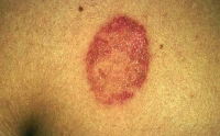
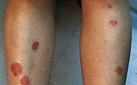
Tinea Corporis
This is an infection of the body. It can be the limbs, the face or the neck. Children are more likely to have this kind of infection.
The infected area will appear as a small red lump initially, which then spread peripherally to become a big round circular patch. Very often only the round margin is seen, with the central area being smooth and flat. It can range from no itch to severe itchy feeling.
|
|
|
Tinea Unguium
This is infection of the nails, and will be discussed under the section Hair & Nails Problems.
How To Avoid Fungal Infection
The most important thing is to keep the skin dry, particularly between fingers, between toes and in the groins. If there is excessive sweating, it should be reduced by anti-perspirants. However, talc powder added with anti-fungal property is more valuable, which can absorb the sweat, prevent skin friction, as well as inhibit the fungi.
Try to shorten the period of wearing shoes, which should be airy and not occlusive. The socks should be changed more often, and can be pre-treated with anti-fungal talc powder before putting on.
Slippers or socks should not be shared with family members who are suffering from fungal infection of the feet or toe nails. Avoiding walking on the floor bare-footed. In the case of fungal infection of the hair and scalp, do not share the combs, hats, pillows and towels.
Try not to touch and scratch any fungal infection of the skin and nails. Otherwise it may be spread to other areas on the body, or to someone else. When it is suspected that the family pet is having fungal infection, take it to see the vet and receive treatment as soon as possible.
Clinical Diagnosis
Usually the doctor can diagnose fungal infection from the clinical features and it does not require any special tests. In cases when it may be confused with other skin diseases, and a definitive diagnosis is needed, then a skin sample can be obtained from scrapping the skin, or a hair sample from cutting some hair off. These are then sent to the laboratory for examination under the microscopy, or culture.
Common skin diseases that can be confused with fungal infection include eczema and psoriasis.
Treatments
For all the cases of tinea in different areas, both topical and oral treatments can be used.
In general, topical cream is used for minor skin infection in small area. Some useful creams are Miconazole, Clotrimazole, Econazole and Terbinafine. Very often they are mixed with steroid to reduce the inflammation and itch, but inappropriate use may lead to more wide spread infection that is more difficult to treat.
For infection of a more extensive area, oral treatment is required. The mostly used are
Terbinafine and Itraconazole. The usual course of treatment is from 2 to 6 weeks. Because of the short period, there are usually no significant side effects. However for people with liver diseases, it is best avoided.
If there is severe skin maceration, such as a severe case of wet tinea pedis, the skin can be soaked with diluted potassium permanganate. It is done once daily for 15 to 20 minutes. It helps to dry up the skin, but it will stain the skin, nails, and clothing with a dark purplish brown colour.
For co-existing bacterial infection, oral antibiotic will be required too. Some creams have mixed preparations of steroid, anti-fungal, and antibiotic, but they are very often abused to treat a variety of skin problems.
Pityriasis Vesicolor
This is a common skin disease caused by an overgrowth of the yeast fungus called Pityrosporum orbiculare (Malassezia furfur) on the skin surface. The growth of these organisms relies on the presence of mature sebaceous (oily) glands; therefore it is not common before puberty, and mainly affects young adults.
Most adults have these organisms on their skin and normally they do not cause any skin problems. However, during the hot and wet seasons, in subtropical and tropical regions, they may grow excessively and result in a harmless skin disease. Most sufferers first notice this problem after the summer, which then spread gradually.
Profuse perspiration and high production of sebum (oil) by the skin make it easier for the fungus to multiply and spread, for instance in teenagers, but the condition is not contagious.
Signs
Pityriasis versicolor is commonly seen on the upper part of the back, the neck, and the chest, but it can be occasionally found on the entire body.
In the beginning, there are only a few small patches, often unnoticed. The growth is very slow and may take a few months before they are noticed.In the summer the infection is seen as well-defined, uneven patches. The patches are pale-red or brownish, and often merge into big irregular blotches. The surface is slightly scaly when lightly scratched with a sharp object. The fine scales are signs of active infection, because when it is treated, they are no longer present.
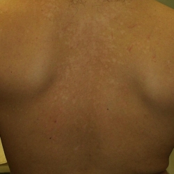
The colour may be unstable and vary from a lighter to a darker hue when compared to the surrounding skin. The organisms may inhibit the formation of melanin pigment, and as a result, the patches are much whiter in colour, and will not tan after exposure to sun, whereas the surrounding will tan as usual. Therefore the contrast is increased and it may be the first time that they are noticed after the person has spent a sunny weekend on the beach. White patches will take a long time to return to normal skin colour even after successful treatment.
In some people, Pityrosporum orbiculare infects the hair follicles on the back and on the chest, which causes highly itchy acne-like spots to appear.This condition is called pityrosporum folliculitis, and it is more common in people who are between 30 and 40 years of age, particularly women.
Diagnosis
Skin scrapings, when treated with special dyes, will show the yeast organisms under microscopy. The patches may show a slight fluorescent yellow when examined under ultra-violet light (Wood's light examination). However, these are usually unnecessary as the signs are quite typical to be recognised.
Treatments
It mainly relies on topical treatments. Shampoo containing selenium sulphide or ketoconazole can be used to wash the area. It is very useful for large affected area. Alternatively it can be put on directly for a few hours every day. The problem with the selenium sulphide shampoo is the unpleasant smell.
Anti-fungal preparations in creams or sprays can be used twice daily for 3 to 4 weeks. In general, oral anti-fungals, such as itraconazole, are not necessary unless in severe cases, or in pityrosporum folliculitis.
The flakiness will disappear after successful treatment, but the whitened patches will not return to normal skin colour straight away. No further treatment is needed, and it will just take a longer time.
The condition is likely to recur, which can be prevented by washing with either selenium sulphide or ketoconazole shampoo.